

“FIRST OF ITS KIND IN THE NATION”
Dedicated Exclusively to Diagnostic and Consultative Pathology of the Oral Cavity, Head and Neck

CLINICAL INFORMATION
Forty-one year old healthy man with a solitary 1.8 cm nodular, firm, ulcerated lesion on the posterior dorsal tongue present for one week. Negative past medical and dental history.
SPECIMEN:
Incisional biopsy of mucosa coated with yellow, soft, pasty material.
FINAL DIAGNOSIS:
Traumatic ulcerative granuloma with stromal eosinophilia and atypical lymphoid infiltrate.
DISCUSSION:
Traumatized ulcers frequently show a mixture of degenerative and regenerative changes, often times displaying highly atypical cells. The epithelium adjacent to the ulcer in this particular biopsy does not show features of concern. The bulk of the mass consists predominantly of large lymphohistiocytoid cells expanding the stroma (mass effect) and splaying striated muscle “fibers. The nuclei are large and pleomorphic and show irregular chromatin distribution and atypical mitoses. Few eosinophils, plasma cells and small mature lymphocytes are present.
In view of the degree of cytologic atypia, a panel of immunohistochemical stains was performed with very worrisome results. Molecular studies ruled against a monoclonal (malignant) process. The above diagnosis was rendered.
Traumatic Eosinophilic Ulcerative Granuloma was originally described by Popoff in 1956 as a deep ulcer on the tongue of adults. It was recognized as a distinct entity in 1970 and since then it has been described in other areas of the oral cavity under a variety of often confusing names. A similar lesion occurs in young children (Riga-Fede disease). The clinical presentation and histomorphology can be alarming, therefore this diagnosis should be made with caution.
In view of the location and microscopy, the possibility of squamous cell carcinoma is slim. Infection and other neoplasias were considered and ruled out. What is different about this case is that the inflammatory component expands the stroma producing a mass. The history of rapid growth militates against developmental lesions such as ectopic tissue, i.e thyroid and raises the possibility of malignancy, thereby the work-up.
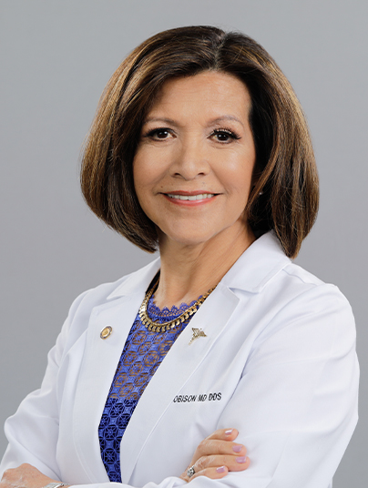
Diagnosis by Rosa H Robison, MD. DDS. FCAP.
Ancillary testing and case discussion performed at the University of Florida Medical Pathology Laboratory, Department of Hematopathology, Gainesville, FL.
A special thank you to Dr. Rigo Cornejo for the opportunity of studying this very challenging
case and for his longtime trust and support.
case and for his longtime trust and support.

- Diplomate of the American Board of Medical
Pathology.
- Diplomate of the American Board of Oral and
Maxillofacial Pathology.
- Fellowship Training: Soft Tissue Neoplasia and
Immunohistochemistry.

5036 Dr. Phillips Blvd. #315 Orlando, FL
T: 407-286-2330 F: 407-523-0496
mddds@ohnlab.com


 Es
Es En
En